
CHEST X-RAY · AI · CT
A case in which an AI finding led to a lung cancer diagnosis
Chest X-ray and detailed assessment with CT
At our clinic, physicians use CXR Finding-i, an AI image-analysis system, to assist their review of chest X-rays. Here we present a case in which an AI finding prompted further investigation, leading to a diagnosis of stage I lung cancer.
Reviewed by Koshiro Nishimoto, Clinic Director
Updated November 9, 2026

and hold.
Select an image to view it at full size.
01
Chest X-ray taken at our clinic
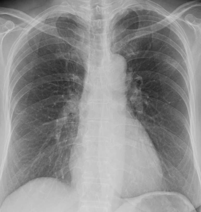
This chest X-ray was taken at our clinic. In this case, the abnormal shadow was subtle, making the lesion difficult to recognize on the chest X-ray alone.
02
AI flagged an abnormal shadow
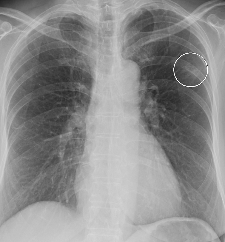
The circle indicates the area where AI flagged an abnormal shadow.
In this case, the lesion would have been difficult to recognize without AI assistance. The AI finding provided an important clue for suspecting lung cancer and proceeding to further investigation.
The physician reviewed the AI result and arranged a chest CT for a closer assessment.
03
Chest CT and the subsequent diagnosis
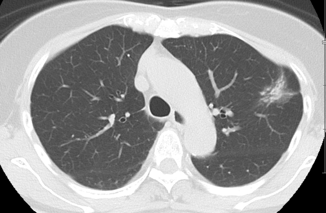
Chest CT provided a more detailed assessment of the lesion in the area identified on the X-ray. After further evaluation, the patient was diagnosed with stage I lung cancer.
This case illustrates how combining physician review with AI assistance can help identify a lesion and lead to a subsequent diagnosis. At our clinic, AI analysis is used as a diagnostic aid to guide further testing when appropriate.
AI supports physician diagnosis; it does not diagnose lung cancer on its own and cannot detect every lesion. This is one case. Physicians determine which tests are needed and make the final diagnosis.